The complexity of the aberration detection and correction process for PALM/STORM microscopy in biological samples has long been a limiting factor for widespread use of adaptive optics in biological imaging. The closed-loop method, though recognized as the best approach in terms of accuracy and speed, is usually difficult to implement due to the absence of a point source for direct wavefront sensing in biological samples, and the addition of fluorescent beads to the sample is seldom possible. Image-based iterative aberration detection algorithms can solve this problem, but its use in PALM/STORM microscopy is still a challenge. Indeed, PALM/STORM super resolution raw images are composed of single molecule detections (point sources), which appear at a different place in every acquired frame, and their intensity varies over time. This means “classical” merit functions, like maximal intensity or contrast, don’t work.
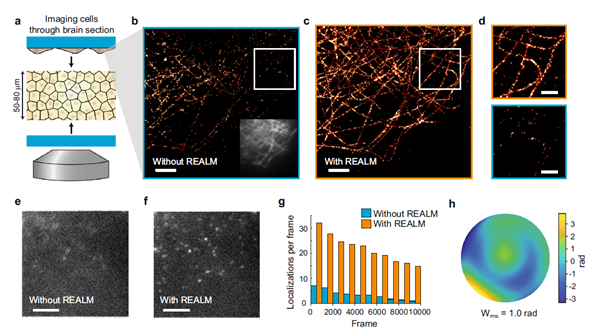
Recent innovations are stretching these limits. A publication in Nature Communications by the group of Lukas Kapitein from Utrecht University reports successful application of a novel merit function based on a Fourier transform of raw images, which, therefore, does not depend on the intensity and location of detections in every frame. Their method, which is called REALM, allows direct use of the “blinking images” of the PALM/STORM technique and permits detection of aberrations on the fly. The authors need as little as about 300 frames to detect aberrations, apply the correction using a deformable mirror and then acquire the PALM/STORM sequence using a perfect PSF. By correcting aberrations, in particular in depth, the number of counts per frame is demonstrated to be increased by a factor of at least 4, due to the restoration of the PSF quality.
Lukas Kapitein’s group is one of the leading teams in the world studying the cytoskeleton of neurons. They use several innovative research methods to understand the mechanisms by which cells establish and maintain their precise shape and intracellular organization. Their REALM technique in particular was made possible thanks to our MicAO 3DSR adaptive optics add-on for super resolution PALM/STORM systems. The method opens the door to deep SMLM in tissue with unprecedented 3D resolution. For even more information about the method, (re)watch the webinar organized by Imagine Optic and presented by Marijn Siemons, PhD student in Lukas’s group, (and including a live demo!).
If you’re interested in finding out more about our AO solutions for Microscopy, you can reach us at sales@imagine-optic.com or through the contact form (red enveloppe on the side).