Adaptive Optics for Immunology and Intravial imaging
Immunology is a science of behaviour: where a cell goes, what it touches, how long the contact lasts. Whether it is to observe interactions within a lymph node, spleen or a tumour, light distortions will be encountered. Adaptive optics for immunology measures and corrects these distortions with a deformable mirror, restoring a sharp, symmetric focus deep inside the specimen. Cells stay segmentable deep in the specimen, contacts stay distinguishable from proximity, and none of it requires more laser poweravoiding to cause more photostress.

Related Products
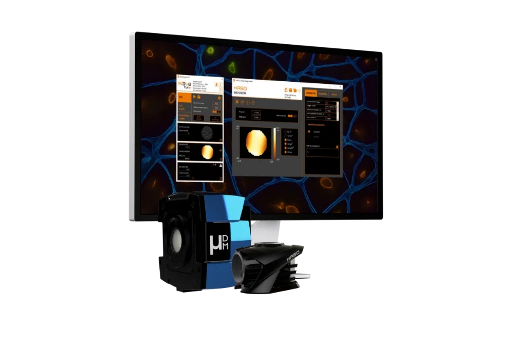
AO KIT BIO
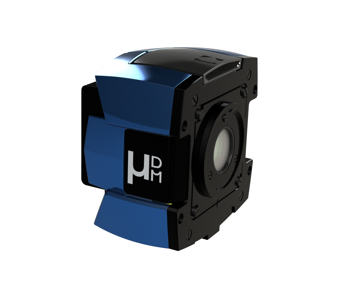
mu-DM
How adaptive optics (AO) can be the solution
Adaptive optics measures the aberrated wavefront and applies its conjugate with a deformable mirror, maintaining a sharp, symmetric point spread function throughout the imaged volume rather than only near the surface.
For immunology the benefit arrives mainly as signal. Because two-photon excitation scales with the square of intensity, a tighter focus recovers fluorescence faster than it recovers resolution — so the first thing correction buys is the signal-to-noise margin that segmentation and tracking algorithms need. Uniform resolution across the volume matters as much as peak resolution: it is what makes contact events at 200 µm comparable with contact events at 50 µm.
The practical result is that you can visualize cellular interactions as events rather than infer them from proximity — resolving the contact interface itself, at the depth where it occurs, with the same fidelity as one imaged near the surface.
That recovered signal allows lower excitation power instead of brighter images, which removes the photostress confound. Correcting the optics rather than raising the power is what allows you to track immune dynamics with adaptive optics without altering the physiology under study.
Intravital microscopy of immune responses
Immune behaviour only exists in intact tissue, so immune cell imaging is built on two-photon intravital microscopy, which reaches into living lymph node, spleen and tumour with enough sectioning to follow single cells over hours. Tissue-induced aberration limits how deep that resolution survives, and therefore how much of the immune response is actually observable.
Imaging lymph nodes, spleen and tumour microenvironments
Each immunological tissue distorts light in its own way, so the aberration an adaptive optics system has to correct differs by organ rather than only by depth.
| Tissue / model | Aberration source | Optical impact | AO benefit |
|---|---|---|---|
| Lymph node | Capsule, densely packed lymphocytes, follicle boundaries | Index steps at the capsule and between cortex and paracortex; cells merge with depth | Keeps tightly packed T and B cells separable in depth |
| Spleen | Red pulp blood content, haemoglobin absorption, trabeculae | Strong absorption combined with refractive index variation | Recovers signal without increasing photostress in absorbing tissue |
| Tumour microenvironment | Dense collagen, necrotic regions, lipid-rich zones, irregular vasculature | Field-dependent aberrations across single imaging planes | Maintains uniform resolution across the tumor |
| Bone marrow (calvarial) | Bone layer overlying the marrow cavity | Severe spherical aberration and defocus at the bone–tissue interface | Restores cellular resolution beneath intact bone |
Main sources of aberration in common immunological tissues, and what adaptive optics recovers in high-resolution immune cell microscopy.
Send us info about tissue and microscope setup
We’ll tell you what adaptive optics can recover in your preparation — tissue, animal model, imaging depth and microscope type are all we need to start.
Request a feasibility checkOvercoming key imaging challenges in live immune dynamics
Living tissue is optically heterogeneous in a way that is difficult to model and impossible to avoid. Refractive-index mismatch between dense cell bodies, collagen fibre networks, adipose regions and vasculature deforms the wavefront, and the focal spot degrades progressively with depth.
The consequence for immunology is specific. When the focus broadens, the boundary between two cells becomes ambiguous, and a contact — the event that defines immune cell communication — can no longer be distinguished from proximity. Studies of macrophage and T cell interactions depend on that distinction being made correctly and consistently across a volume.
Motion makes this harder. High-definition T cell tracking requires linking the same cell across successive frames, which fails when signal-to-noise drops below the threshold the tracking algorithm needs. Segmentation errors do not distribute randomly; they accumulate at depth, where the aberration is worst, and bias the velocity and contact-duration statistics derived from the dataset.
The standard remedy is more excitation power, and here it is worse than usual. Photostress alters immune cell activation state and migration velocity. Raising power to see the behaviour more clearly changes the behaviour being measured.
Differentiating true membrane contact from proximity
Overlapping Point Spread Functions (PSFs) at depth blur cellular boundaries, causing adjacent cells to merge into a single signal. This introduces false positives when analyzing T cell-macrophage interactions or immune synapses. AO restores a diffraction-limited PSF, allowing researchers to quantify cell-to-cell contact with similar criteria at 200 µm as at 50 µm depth.
Label-Free matrix imaging with Second-Harmonic Generation (SHG)
SHG visualizes extracellular collagen scaffolds alongside migrating immune cells. As a coherent optical process, SHG signal intensity drops faster than incoherent fluorescence under wavefront error. AO recovers SHG contrast disproportionately, enabling accurate registration of cell tracks against the structural matrix.
Mitigating photostress & physiological artifacts
Increasing laser power to compensate for signal loss alters immune activation states and migration velocities. By concentrating photons at the focal point, AO boosts fluorescence signal while keeping excitation intensity low enough to preserve natural immune behavior.
Handling tissue motion (breathing, heartbeat, blood Flow)
- What AO does : Corrects phase aberrations caused by refractive index variations.
- What AO does not do : Correct physical tissue displacement (which requires mechanical stabilization, gating, or post-hoc registration).
- Speed Requirement: Live intravital imaging demands fast wavefront measurement routines so correction occurs before the tissue structure shifts.
Adaptive optics solutions for intravital microscopes
µDM
Deformable mirror for microscopy
For labs that build or modify their own intravital system. µDM sits conjugate to the objective pupil and runs in closed loop with a wavefront sensor or in open loop for sensorless correction. Linear response with no hysteresis keeps optimisation converging quickly when the correction has to be re-found each time the field moves, and temporal drift compensation holds it across a multi-hour timelapse.
AO Kit Bio
Complete adaptive optics kit
For labs that want sensor-based correction without designing the system from scratch. The kit combines a deformable mirror, a Shack-Hartmann wavefront sensor and control software, ready to integrate into the excitation path of a custom two-photon microscope. Extended-scene wavefront sensing means no guide star is needed in scattering tissue.
Not sure which fits your setup? Our application team designs the optical integration with you.
Request a feasibility checkFrequently asked questions: adaptive optics for live immune dynamics
Can adaptive optics help image T cells inside a lymph node in vivo?
Yes, and the lymph node is one of the clearest cases for it. Lymphocytes are packed tightly enough that an aberrated focus merges neighbouring cells, which is what breaks tracking in the deep paracortex. Correcting the wavefront keeps individual cells separable, so migration paths and contact events can be followed where the response is actually organised.
Does adaptive optics help detect immune synapses?
– Live Intravital Imaging : Yes. AO provides the spatial resolution needed to distinguish true membrane contact from close cell proximity.
– Sub-Cellular Architecture : For nanoscale receptor microclusters on fixed cells, single-molecule localization microscopy (e.g., MICAO 3DSR) restores 3D PSF symmetry.
Is adaptive optics compatible with multicolour intravital imaging?
Yes. AO actively compensates for chromatic focal shifts between color channels, ensuring precise spatial colocalization across excitation wavelengths.
How does adaptive optics cope with tissue motion?
It does not correct motion. Adaptive optics corrects phase aberration; breathing, heartbeat and blood flow are displacements, and they are handled by stabilisation, gating and image registration. What motion does affect is how quickly the correction must be measured, since a wavefront measured before the tissue moves is no longer the right one afterwards.
What imaging depth can be reached in a lymph node with adaptive optics?
There is no single figure, because depth depends on the animal model, the labelling, the excitation wavelength and the objective. Adaptive optics extends the usable depth wherever aberration rather than scattering is the limiting factor. Once scattering dominates, correction no longer helps, and that crossover is what sets the practical limit in any given preparation.
Planning to add adaptive optics to image live immune dynamics ?
Our microscopy team will review your setup and recommend implementations.
Book a call with our microscopy team Download the datasheetsBlog Posts / Application Notes
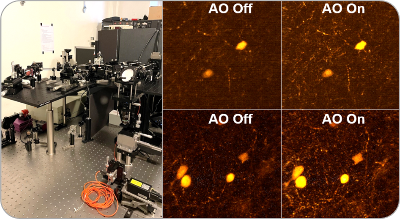
One more prototype of a microscope with adaptive optics: this time for 2-photons!

Lattice Light-Sheet with Adaptive Optics
Related Applications
Developmental biology

Drug Development
Neuroscience