A custom-built 2-photon microscope incorporating a new, fast adaptive optics (AO) approach now provides its first AO-enhanced images.
When targeting high-resolution imaging of biological samples at large depths, non-linear microscopy, and in particular 2- or even 3-photon fluorescence microscopy, is usually a technique of choice. Due to near-infrared illumination and intrinsic optical sectioning capability, multiphoton microscopy provides deep 3D imaging with virtually no background and a great versatility enabled by the use of 2D scanning in the illumination path. This method is now widely employed for neuroimaging, particularly in scattering media such as rodent brain. However, as for all high-resolution optical microscopy techniques, its performance is severely reduced as a consequence of optical aberrations induced by the sample, especially in depth, which strongly degrades the illumination point spread function (PSF) resulting in a significant loss of signal and contrast.
Recently, we demonstrated a new, fast and simple AO approach, as well as its integration in a light-sheet microscope (more details here). This new AO method is based on direct wavefront sensing without the need for a guide star, enabling both fast correction (typ. 1 to 5 s) and reduction of the constraints of use. Aiming to provide users with an easy operation of AO in multiple microscopy modalities, Imagine Optic, together with a team of researchers from Ecole Supérieure de Physique et de Chimie Industrielle (ESPCI, France) and Ecole Normale Supérieure (ENS, France), adapted AO to the excitation path of a custom-built two photon microscope. This wavefront sensing approach was demonstrated to be particularly efficient in scattering conditions (publication here).
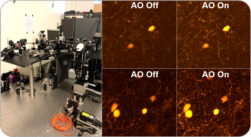
Our prototype microscope is using one of Mirao line of large stroke, high stability deformable mirrors and allowed us to acquire first images of ex vivo samples mainly consisting in fixed fluorescent mouse brain slices, at depths reaching 200µm (see upper figure, representing a maximum projection over 10µm brain slice). Only 4 iterations of the closed-loop optimization, corresponding to 2-3 seconds, were necessary to achieve this aberration correction. AO correction enabled an average three times increase of the signal, providing sharper morphological details such as axons or dendrites. These latest results have been recently presented at the SFO congress in Nice (France): Imperato S. et al., Extended scene adaptive optics for 2 photon Neuroimaging in the mouse brain, 05 Jul. 2022. They offer a great promise, specifically regarding the next steps of in vivo recording of functional signals, as well as product development.
These results have been achieved in the frame of the INOVAO project (funding Agence Nationale de la Recherche, ANR-18-CE19-0002).
#AdaptiveOptics #Microscopy