Adaptive Optics for Drug Screening and High-Content Imaging
A phenotypic screen is only as reliable as the segmentation underneath it, and segmentation is decided by the optics before any analysis runs. Plate bottoms, immersion mismatch and thick 3D cultures all distort the wavefront, and they distort it differently from one well to the next. Adaptive optics for drug screening corrects that distortion in hardware, per well, under machine control. Object boundaries stay defined across the plate, morphological measurements stay comparable, and the excitation dose can come down rather than up.

Related Products
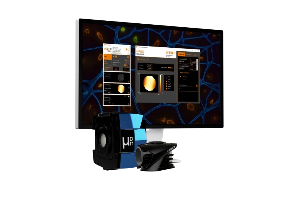
AO KIT BIO
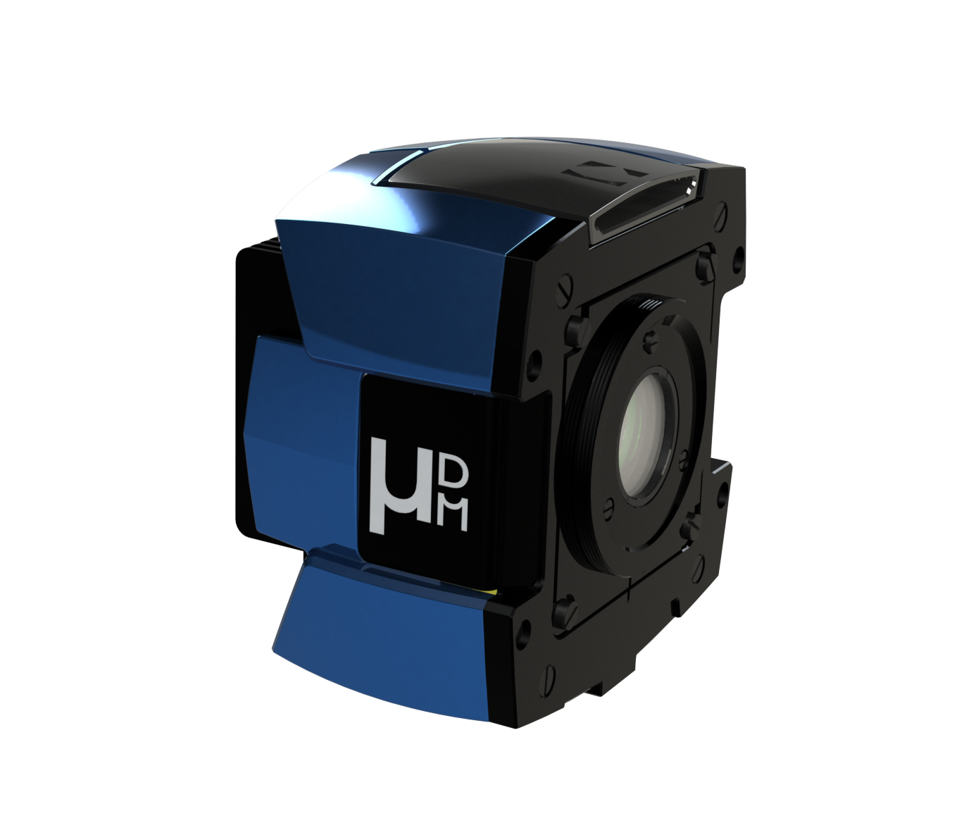
mu-DM
Adaptive optics for high-content screening (HCS)
High-content platforms quantify cellular morphology, localization, and target engagement across compound libraries. Optical aberrations degrade image fidelity prior to segmentation, turning physical artifacts into false biological hits.
Imaging 3D spheroids and organoids in multiwell plates
Screening has moved toward 3D culture because spheroids and organoids predict in vivo response better than monolayers. Optically they are far harder: each object is hundreds of micrometres of densely packed tissue sitting in a refractive-index-mismatched medium, so the aberration varies with depth into the object and between objects in the same well.
| Sample format | Aberration source | Optical impact | AO benefit |
|---|---|---|---|
| 2D monolayer (glass-bottom) | Coverslip thickness tolerance (well-to-well) | Spherical aberration varying across the plate | Uniform focus qualityacross the plate |
| 2D monolayer (plastic-bottom) | Polymer bottom thickness and birefringence | Stronger spherical aberration than glass, worst at well edges | Recovers resolution without moving to costlier glass plates |
| Spheroids | Dense cell packing, medium refractive-index mismatch | Rapid signal drop with depth into the spheroid | Restores internal focus where segmentation typically fails |
| Organoids | Heterogeneous architecture, lumens, lipid-rich regions | Aberration varies within a single object, not only with depth | Per-field correction keeps morphology measurable throughout |
| Matrix-embedded cultures | Hydrogel or basement-membrane extract index mismatch | Strong defocus and spherical aberration with gel depth | Large-stroke correction holds the target focal plane |
Main sources of aberration in common screening formats, and what adaptive optics recovers.
Send us your assay and imaging platform
We'll tell you what adaptive optics can recover in your screen – assay, plate format, culture type and imaging platform are all we need to start.
Overcoming Optical Bottlenecks in Automated Platforms
Plate-induced aberrations and their impact on screening data
Microscope objectives are calibrated for a specific coverslip thickness and immersion medium. Multiwell plates violate these parameters inconsistently due to:
- Bottom thickness tolerances : Micron-level variations across a plate introduce depth- and NA-dependent spherical aberration.
- Polymer substrates : Plastic bottoms differ from glass in refractive index and introduce birefringence.
- Meniscus and evaporation : Uneven medium depth across wells alters the optical path during long automated runs.
Spinning-Disk Confocal Throughput and Signal
Spinning-disk systems rely on fluorescence returning through excitation pinholes. When wavefront errors broaden the emission Point Spread Function (PSF), in-focus signal is rejected by the pinhole array. AO restores pinhole coupling, recovering both optical sectioning and fluorescence intensity simultaneously
Uncorrected aberrations create structured noise, degrading images worse at well edges or deep inside organoids. This biases downstream metrics derived from segmentation masks (nuclear area, cytoplasm-to-nucleus ratio, texture features). Hardware wavefront correction restores true object boundaries at acquisition, ensuring a documented instrument state for regulated workflows.
Protecting Data Integrity and Segmentation Masks
Uncorrected aberrations create structured noise, degrading images worse at well edges or deep inside organoids. This biases downstream metrics derived from segmentation masks (nuclear area, cytoplasm-to-nucleus ratio, texture features). Hardware wavefront correction restores true object boundaries at acquisition, ensuring a documented instrument state for regulated workflows.
Lowering Excitation Dose for Live-Cell Toxicity Assays
Compensating for signal loss with higher laser power induces photostress, altering cellular baselines during viability and toxicity screens. By concentrating available photons at the focal plane, AO achieves high Signal-to-Noise Ratios (SNR) at lower illumination intensities.
Adaptive optics solutions for screening platforms
µDM
Deformable mirror for automated imaging
The corrector specified for screening. Trigger-in and trigger-out synchronise correction to the acquisition sequence, so a new wavefront is applied per well or per plate under machine control. Linearity above 99.5% with no hysteresis means a commanded shape is the applied shape on every cycle, which is the reproducibility requirement that separates a screening instrument from a research microscope. Temporal drift compensation holds performance across long plate runs, and embedded electronics with a single USB3 connection keep integration simple inside an enclosed instrument.
AO Kit Bio
Complete adaptive optics kit
Supplies the rest of the loop: a HASO4 wavefront sensor, WaveTune software, and sensorless 3N and phase-diversity algorithms that derive the correction from the acquired image with no reference bead and no operator. For platform integration the relevant deliverable is WaveKit Bio, a C++ and Python SDK that places the correction loop inside your own control software rather than alongside it.
Building or modifying a screening platform? Our application team designs the optical integration with you.
Request a feasibility checkPlanning to add adaptive optics to a screening platform?
Our microscopy team will review your setup and recommend implementations.
Book a call with our microscopy team Download the datasheetsFrequently asked questions: adaptive optics in high-content screening.
Can adaptive optics run automatically in a high-content screening workflow?
Yes. Sensorless, image-based correction derives the wavefront from the acquired image itself, with no reference bead and no manual input. mu-DM’s trigger-in and trigger-out let the acquisition system decide when a new correction is applied — per well, per plate or per field — so the loop runs under machine control rather than operator control.
How long does correction add per well?
The overhead is the measurement step, not the imaging: once a correction is found it is held on the mirror while images are acquired at full speed. How long the measurement takes depends on the algorithm and how many modes are optimised, and at plate scale the usual approach is to re-measure per plate or per region rather than per well. We size this against your throughput during a feasibility check.
Does adaptive optics improve imaging of 3D organoids in plates?
This is where the aberration is most severe, so it is where correction gives the most back. A thick 3D culture introduces substantial refractive-index mismatch, and the focal spot degrades quickly with depth into the object. Correcting it recovers signal and boundary definition inside the organoid, which is exactly where segmentation of a 3D culture breaks down.
Does adaptive optics improve assay quality metrics such as the Z′-factor?
It acts on the terms the Z′-factor is built from, though we have not measured it directly. Z′ depends on the separation between positive and negative controls and on the variability within each. Correction raises signal-to-noise, which widens the separation, and it removes a source of well-to-well optical variation, which narrows the spread. Whether that moves Z′ enough to matter depends on how much of your assay variance is optical rather than biological or dispensing-related — which is worth measuring on your own plates before assuming either way.
Is adaptive optics compatible with commercial high-content imagers?
Not as a retrofit to a closed commercial platform. Imagine Optic supplies adaptive optics as components — deformable mirrors, wavefront sensors and control software — for integration by instrument builders and by facilities running semi-custom or open systems. If you are developing a screening platform, or your core runs a modifiable instrument, that integration is the conversation to have with our application engineers
Blog Posts / Application Notes

Related Applications
Developmental biology

Immunology
Neuroscience