Adaptive Optics for Developmental Biology
Developing embryos, organoids and whole model organisms are among the most optically demanding specimens in biology. They are thick, they scatter light, and their refractive-index structure changes as tissues form. Adaptive optics for developmental biology measures and corrects the resulting wavefront distortions with a deformable mirror. It restores sharp optical sectioning and single-cell resolution deep inside living specimens, and it does so without raising excitation power or limiting the duration of time-lapse experiments.

Related Products
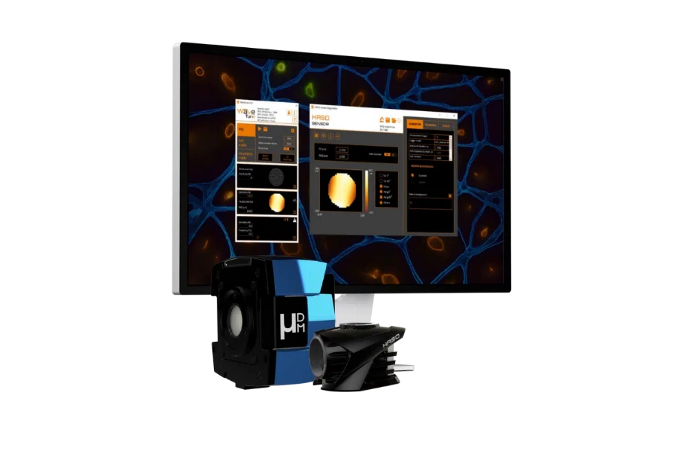
AO KIT BIO

MICAO 3DSR

mu-DM
Light-sheet microscopy for embryo imaging
Studying development means watching an organism build itself. Light-sheet microscopy is the standard choice for embryo imaging, but scattering and refractive-index variation degrade optical sectioning as specimens grow.
Which model organisms benefit from Adaptive Optics?
| Organism / Model | Aberration source | Optical impact | AO benefit |
|---|---|---|---|
| Zebrafish | Yolk, eyes, pigmented cells | Accumulative phase distortion per tissue layer | Recovers deep structural details without changing sample prep |
| Drosophila | Yolk granules, dense lipid droplets | Rapid loss of image quality below outer cell layers | Preserves single-cell focus deep in the embryo |
| Mouse embryos | High tissue thickness, opacity | Severe scattering and wavefront deformation | Enables deep organogenesis tracking |
Main sources of aberration in common developmental biology models, and what adaptive optics recovers.
Why embryos distort the light sheet
- Internal refractive index contrast : Yolk, lipid droplets, and dense nuclei refract light differently from the surrounding cytoplasm.
- Embryo geometry : Curved tissue surfaces act as weak lenses, shifting and tilting the light sheet.
- Mounting media mismatch : Index mismatches from agarose, FEP tubes, or immersion media introduce depth-dependent spherical aberration.
Multi-view and isotropic imaging of whole embryos
Multi-view light-sheet microscopy images the embryo from several angles and fuses the views. This gives near-isotropic resolution and covers the side of the specimen that faces away from the detection objective. Fusion and multi-view deconvolution assume that every view is sharp and that the views can be registered precisely. In practice, each view crosses different tissue, so each one carries different aberrations, and the mismatched point spread functions degrade both registration and deconvolution. Correcting each view with adaptive optics brings the point spread functions back into line. The fused volume is then sharper, and registration errors stop showing up as artefacts in cell positions
Imaging gastrulation and organogenesis deep in the embryo
Cell movement during gastrulation and internal structure formation (heart, neural tube, somites) occurs deep within thick tissue. AO maintains sharp focus across all depth layers, ensuring cell shapes, divisions, and migration paths are quantified with uniform precision.
Send us your sample and microscope setup
We'll tell you what adaptive optics can recover in your embryos – organism, stage, imaging depth and microscope type are all we need to start.
Adaptive optics correction for long time-lapse imaging
A deformable mirror measures and corrects the wavefront and restores the designed point spread function. Most of the aberration is low-order, so stroke matters more than actuator count. The correction is updated as the embryo develops, using sensorless image-based methods, and the recovered signal allows lower laser power.
Cell tracking and lineage reconstruction with a corrected focus
Lineage reconstruction depends on segmenting every nucleus in every frame and linking it across time. When aberrations blur neighbouring nuclei together, segmentation merges them, tracks break, and an error at one time point spreads through the whole lineage tree. Deep cells are affected most, so the resulting lineages are biased towards superficial tissue. With adaptive optics, nuclei stay separable at depth. Tracking software makes fewer errors, there is less manual correction to do, and lineages include the internal tissues that matter most in organogenesis.
Correcting both illumination and detection in a light-sheet microscope
A light-sheet microscope has two optical paths at right angles, and each crosses different tissue. Aberrations in the detection path blur the image. They are corrected by a deformable mirror placed in a plane conjugate to the detection objective’s pupil. Aberrations in the illumination path thicken or bend the light sheet and move it away from the focal plane, which costs optical sectioning. This is handled by light-sheet autofocus and illumination correction. Malivert et al. (2022), a study co-authored by Imagine Optic, combined light-sheet autofocus with image-based adaptive optics in a lattice light-sheet microscope. They obtained fast and robust aberration correction in thick samples.
Adaptive optics solutions for light-sheet microscopes
µDM
Deformable mirror for microscopy
For labs that build or modify their own light-sheet microscope. µDM sits in the detection path, conjugate to the objective pupil, and runs in open loop for sensorless image-based correction or in closed loop with a wavefront sensor. Its large stroke handles the defocus and spherical aberration that dominate in embryos; linear response and embedded electronics make integration simple.
AO Kit Bio
Complete adaptive optics kit
For labs that want sensor-based correction without designing the system from scratch. The kit combines a deformable mirror, a Shack-Hartmann wavefront sensor and control software, ready to integrate into custom light-sheet setups.
Not sure which fits your setup? Our application team designs the optical integration with you.
Request a feasibility checkFrequently asked questions: adaptive optics for embryo imaging
How deep can adaptive optics image into a zebrafish embryo?
There is no single figure, because depth depends on the stage, the region and the wavelength. Adaptive optics extends the usable depth by restoring resolution where aberrations, not scattering, are the limiting factor.
Does adaptive optics slow down light-sheet acquisition?
Only slightly. The correction is measured once for a region or time point and then held on the deformable mirror while images are acquired at full speed. The overhead comes from the measurement step, not the imaging.
How often should the correction be updated during a time-lapse?
It depends on how quickly the embryo changes. A common approach is to re-measure at each developmental stage, or whenever an image-quality metric falls below a threshold, rather than at every time point.
Can adaptive optics be added to an open-source light-sheet microscope?
Yes. Component-level systems such as AO Kit Bio and µDM integrate into custom and open-hardware light-sheet setups. The deformable mirror is placed in a plane conjugate to the objective pupil using a relay, and Imagine Optic can help with the optical design.
Does adaptive optics correct scattering in embryos?
No. Adaptive optics corrects phase distortions, which shift and blur the focus. Scattering removes light from the focus altogether. Adaptive optics recovers the resolution lost to aberrations, and the remaining depth limit is then set by scattering.
Planning to add adaptive optics for embryo imaging?
Our microscopy team will review your setup and recommend implementations.
Book a call with our microscopy team Download the datasheetsBlog Posts / Application Notes

Lattice Light-Sheet with Adaptive Optics: precise and robust aberration correction in thick samples.
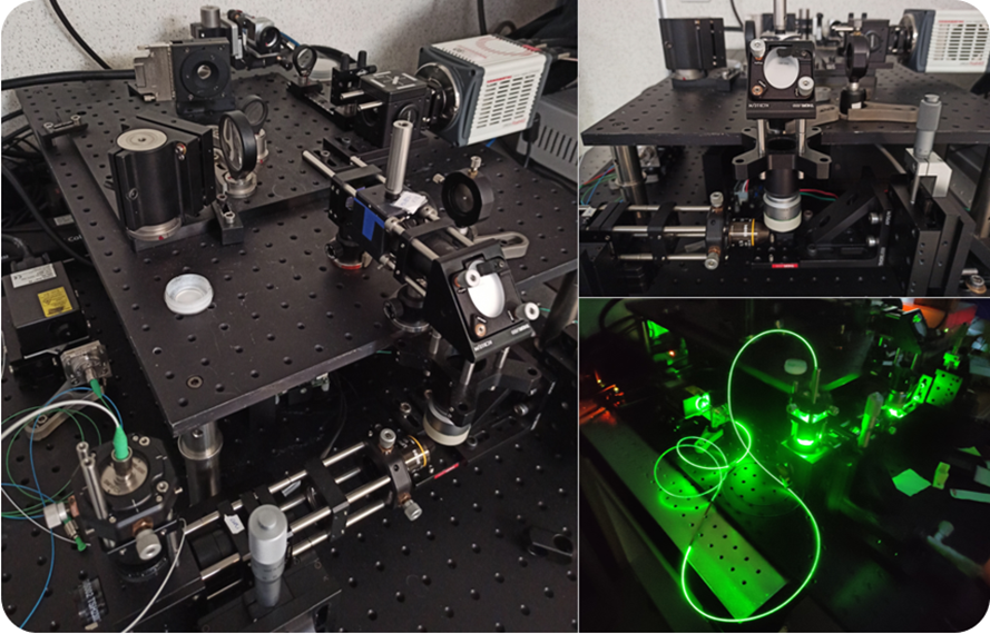
Building a fast adaptive optics light-sheet microscope: first light !
Related Applications

Drug Development

Immunology
Neuroscience