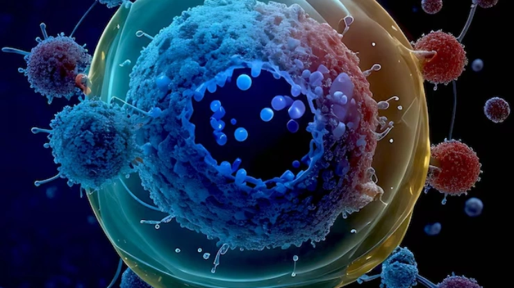
Adaptive Optics for Immunology
Imaging approaches for immunology
Immunology is a discipline about behaviour. Where a cell goes, who it contacts, how long the contact lasts and what passes between the two — these are the measurements, and none of them survives being inferred from fixed tissue.
Intravital and explant immune cell imaging is therefore built on multiphoton microscopy, which reaches into intact lymph node, tumour and mucosal tissue with sufficient sectioning to follow individual cells over time. High-resolution immune cell microscopy inside intact tissue is what lets you decide whether two cells are interacting or merely adjacent. Tissue-induced scattering and aberration limit exactly that judgement.

Challenges of imaging immune dynamics
Living tissue is optically heterogeneous in a way that is difficult to model and impossible to avoid. Refractive-index mismatch between dense cell bodies, collagen fibre networks, adipose regions and vasculature deforms the wavefront, and the focal spot degrades progressively with depth.
The consequence for immunology is specific. When the focus broadens, the boundary between two cells becomes ambiguous, and a contact — the event that defines immune cell communication — can no longer be distinguished from proximity. Studies of macrophage and T cell interactions depend on that distinction being made correctly and consistently across a volume.
Motion makes this harder. High-definition T cell tracking requires linking the same cell across successive frames, which fails when signal-to-noise drops below the threshold the tracking algorithm needs. Segmentation errors do not distribute randomly; they accumulate at depth, where the aberration is worst, and bias the velocity and contact-duration statistics derived from the dataset.
The standard remedy is more excitation power, and here it is worse than usual. Photostress alters immune cell activation state and migration velocity. Raising power to see the behaviour more clearly changes the behaviour being measured.
How adaptive optics can be the solution
Adaptive optics measures the aberrated wavefront and applies its conjugate with a deformable mirror, maintaining a sharp, symmetric point spread function throughout the imaged volume rather than only near the surface.
For immunology the benefit arrives mainly as signal. Because two-photon excitation scales with the square of intensity, a tighter focus recovers fluorescence faster than it recovers resolution — so the first thing correction buys is the signal-to-noise margin that segmentation and tracking algorithms need. Uniform resolution across the volume matters as much as peak resolution: it is what makes contact events at 200 µm comparable with contact events at 50 µm.
The practical result is that you can visualize cellular interactions as events rather than infer them from proximity — resolving the contact interface itself, at the depth where it occurs, with the same fidelity as one imaged near the surface.
That recovered signal can be spent on lower excitation power instead of brighter images, which removes the photostress confound. Correcting the optics rather than raising the power is what allows you to track immune dynamics with adaptive optics without altering the physiology under study.
Related Products
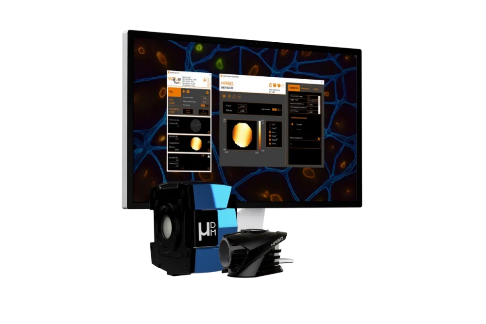
AO KIT BIO
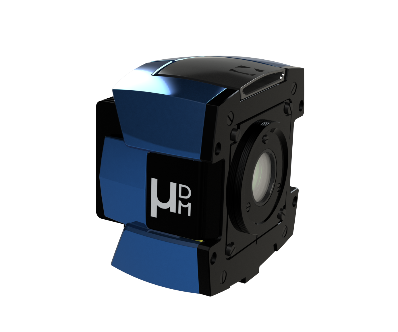
mu-DM
Blog Posts / Application Notes

Related Markets
Developmental biology

Drug Development
Neuroscience